Natural Selections
The Water Bear Solution
By harnessing an ancient animal's superpower, CALS scientists find a way to enhance the clarity of advanced imaging technology.
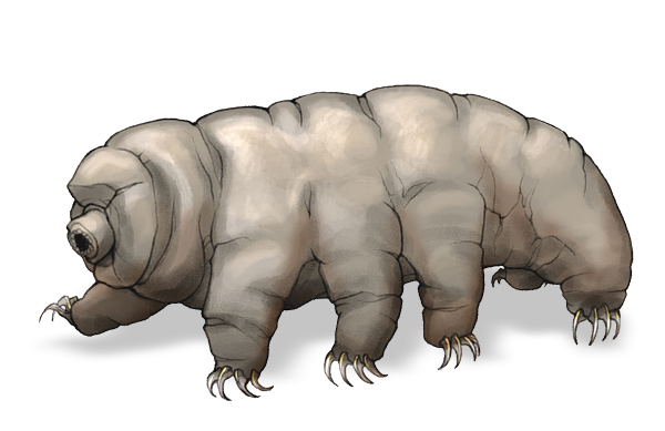
Water bears are an ancient group of microscopic animals known for their pudgy, ursine appearance and their uncanny ability to survive under extreme conditions. The molecules underlying this behavior are helping researchers use modern technology to uncover the basic forms and functions of life’s building blocks.
Also known as tardigrades, water bears are aquatic microorganisms that can endure blistering heat and severe cold thanks to a set of specialized molecules they produce that protect them from damage. These molecules, called late embryogenesis abundant (LEA) proteins, keep the animals’ essential cellular structures and proteins intact, allowing water bears to dry up and go dormant when faced with an uninhabitable environment and then rehydrate and reanimate when it is safe to do so — sometimes decades later. (See Seven Things Everyone Should Know About . . . Tardigrades, Grow, spring 2021.)
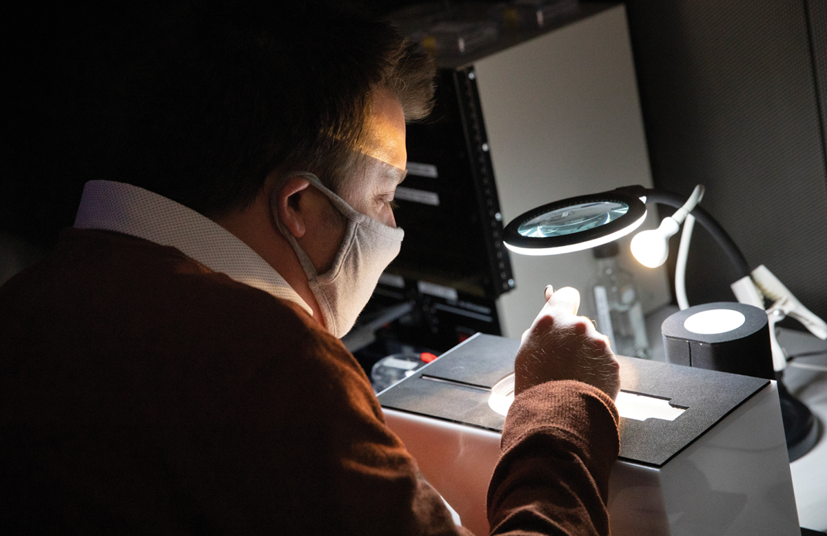
In a study published in September 2024 in Nature Communications, a team of scientists led by biochemistry professor Ci Ji Lim has found that these same proteins can help solve a major technological challenge when it comes to getting high-quality microscope images of a diversity of other cellular structures and proteins using a technique called cryogenic electron microscopy, or cryo-EM.
The study was supported by funding from the National Institutes of Health, the Wisconsin Alumni Research Foundation (WARF), and the UW. It’s one of several new methods of cryo-EM sample preparation developed at UW and licensed by WARF.
Cryo-EM captures miniscule proteins and other biomolecules at a moment in time by freezing samples in a thin, water-based film before taking images of them using a powerful electron microscope. However, around the edges of a sample, where air meets the water, damage to biomolecules is more likely, making it difficult to clearly determine the structures of some proteins.
The technique is important to Lim’s research because he seeks to uncover the relationships between a protein’s form (what it looks like) and function (what it does). Cryo-EM also plays a role in helping identify potential targets for drug therapies. Both require capturing clear images.
Lim wondered whether adding LEA proteins from tardigrades and other microorganisms, such as nematodes, to their microscope samples prior to cryo-EM could prevent damage to the biomolecules and yield better images.
“A lot of times, the protein is attracted to the air-water interface,” Lim explains. “When proteins interact with this interface, they can clump together along the edges or begin to unfold and irreversibly change their form. There are many proteins that we just haven’t been able to fully explore the structures of because of this problem. So, I thought that maybe the LEA proteins, which help proteins in water bears remain robust under challenging conditions, could be harnessed to improve robustness in other contexts.”
Lim and his research team — led by graduate student Kaitlyn Abe — added LEA proteins to samples containing Polα-primase, a protein that is sensitive to the air-water interface. The team also used LEA proteins to image another protein, PRC2, whose structure has been difficult to capture.
The researchers demonstrated that adding LEA proteins is a cost-effective and efficient way to yield clearer cryo-EM images. Other methods to mitigate damage along the air-water interface can be expensive and laborious and can require scientists to use higher concentrations of the molecules they’re studying. LEA proteins allow scientists to use sample concentrations in line with those used with standard sample preparation methods. They could also be used in combination with other protection methods.
“Adding LEA proteins is a readily deployable and cost-effective solution to a bottleneck in cryo-EM research,” Lim says. “It is exciting that we got here using proteins that naturally evolved to do a similar job.”
Scientists are now trying to better understand how these proteins exert their protective properties.
“What is interesting,” says Tim Grant, a professor in the Department of Biochemistry who worked with Lim on the study, “is that even with the addition of the LEA proteins, we’re still seeing biomolecules along the air-water interface, but they’re not unfolding, denaturing, and falling apart. In the presence of LEA proteins, it’s possible to solve structures for proteins that would have just fallen apart before.”
Grant, who is also an investigator at UW’s Morgridge Institute for Research, expects the relationships between biological samples, LEA proteins, and the air-water interface will become sharper as more researchers incorporate LEA proteins into sample processing.
“The air-water interface is a real problem, and water bears have offered a pretty cool solution to resolving that problem,” he says.
Lim looks forward to seeing once-tabled research projects reinvigorated. “Now, there’s new hope for exploring protein structures that we couldn’t before,” he explains. “We’re expanding the toolbox available to scientists for cryo-EM sample preparation, and there’s the potential for us to learn so much more about the structural biology of many proteins that had been challenging to visualize.”
This article was posted in Basic Science, Natural Selections, Summer 2025 and tagged Biochemistry, Ci Ji Lim, cryo-EM, cryogenic electron microscopy, embryogenesis abundant proteins, tardigrades, Tim Grant, water bears.